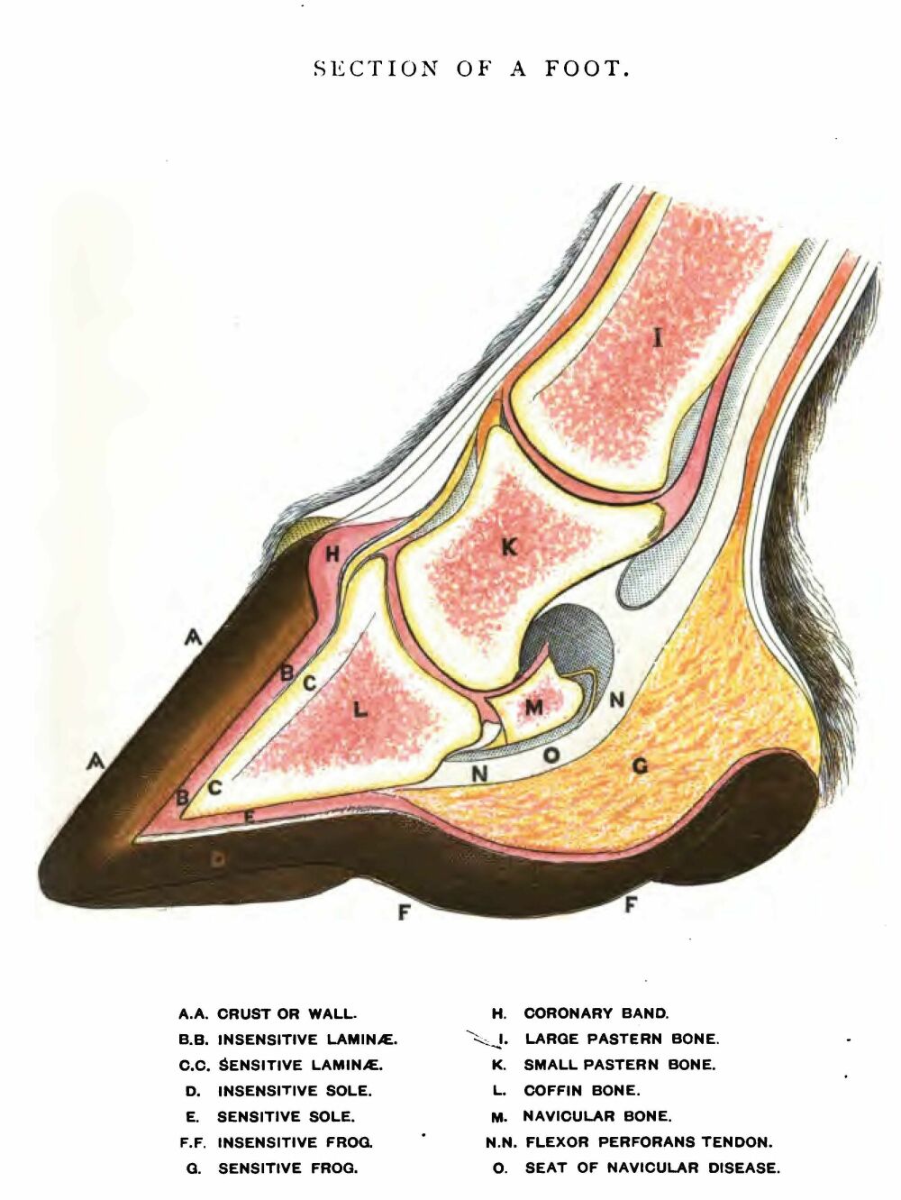
HOOFsmart · anatomy normal hoof cross section drawing labeled
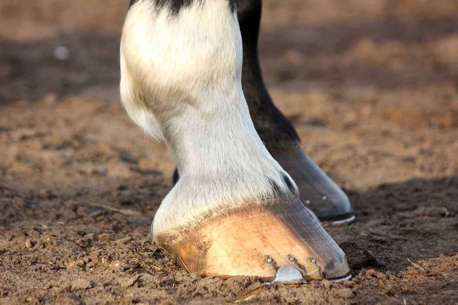
Hoof anatomy. The equine hoof is a unique structure which bears a lot of weight over a small surface area. The term 'no foot, no horse' is extremely important as issues with the hoof can cause major health and movement issues. Last reviewed: 2nd February 2023. The hoof is a complex makeup of structures built to withstand tremendous forces.
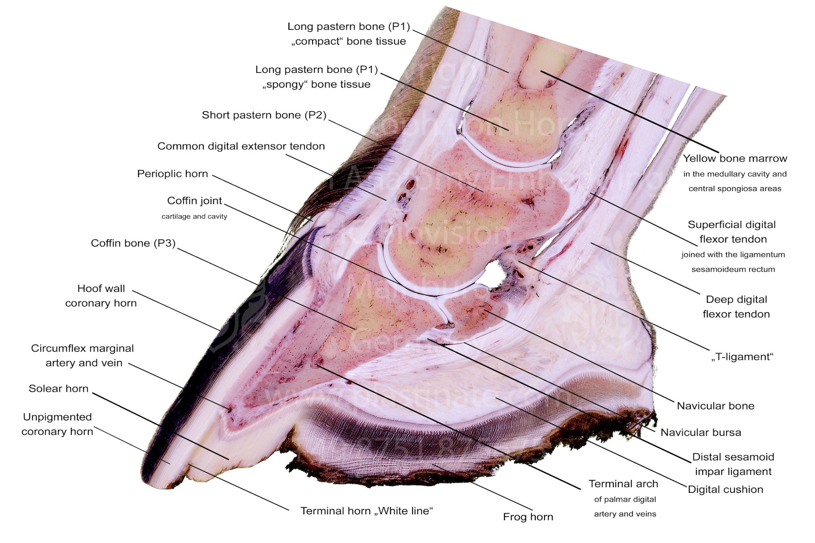
Laminated hoof anatomy chart print Plastination Anatomy Embedding
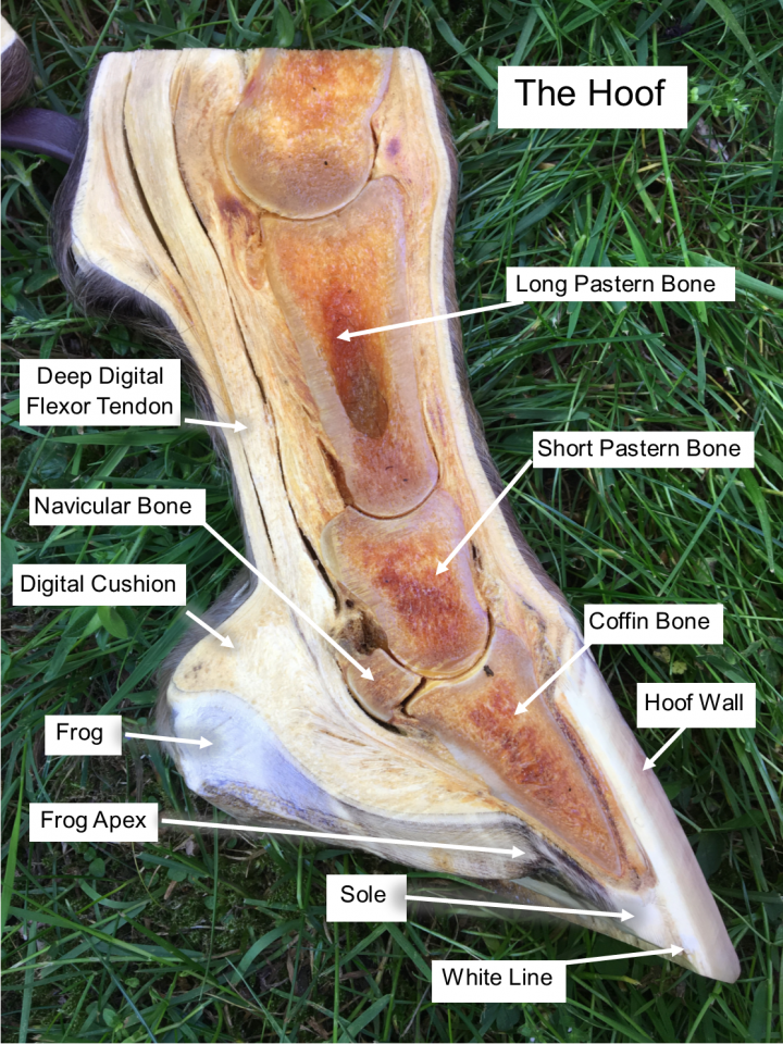
The hoof wall is a weight-bearing structure that grows from the coronet band. It's the exterior-most portion and the part of the hoof that you see when you look at a horse's foot. It is made of keratin, similar to a human's fingernail, and has a low moisture content, making it hard. The wall is essential because it protects the vulnerable.
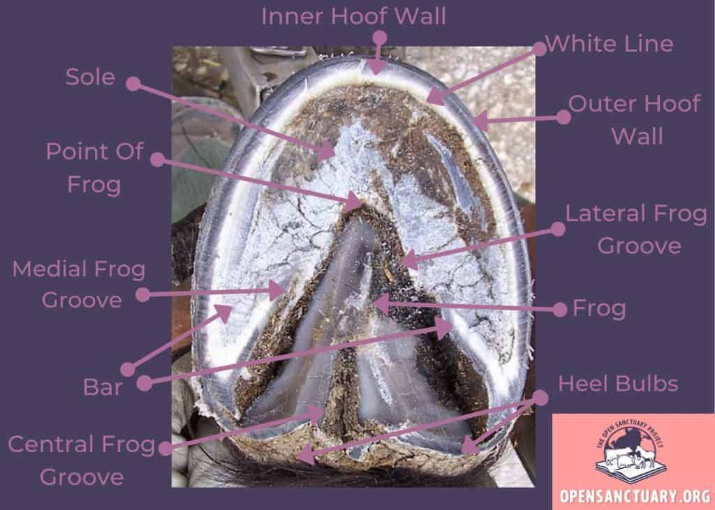
Horse Anatomy The Hoof The Open Sanctuary Project
A horse hoof is the lower extremity of each leg of a horse, the part that makes contact with the ground and carries the weight of the animal. It is both hard and flexible.. Anatomy Transitioning barefoot hoof, from below. Details: (1) periople, (2) bulb, (3) frog, (4) central sulcus, (5) collateral groove, (6) heel, (7) bar, (8.

The Anatomy of the Hoof Hoofcount
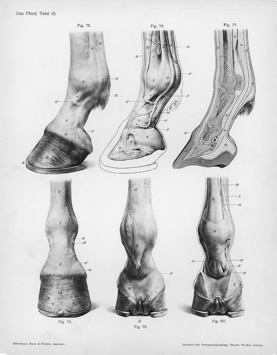
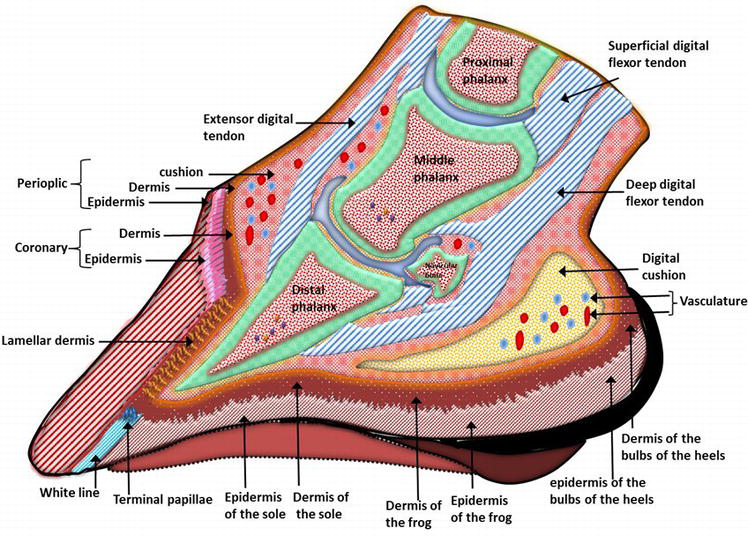
4. The "foot" of ungulates is generally defined as the epidermal hoof capsule and all the tissues and structures enveloped by the capsule, including dermis, subcutaneous tissue, neurovascular tissues, bone, synovial spaces, tendon, ligament, and cartilage. The tremendous weight-bearing forces transmitted through the 4 digits of the horse.
Horse hoof anatomy teaching chart sectional anatomical equine Etsy
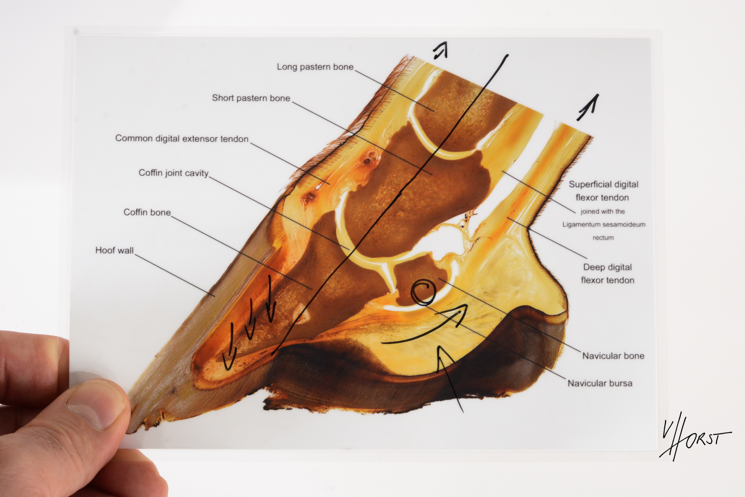
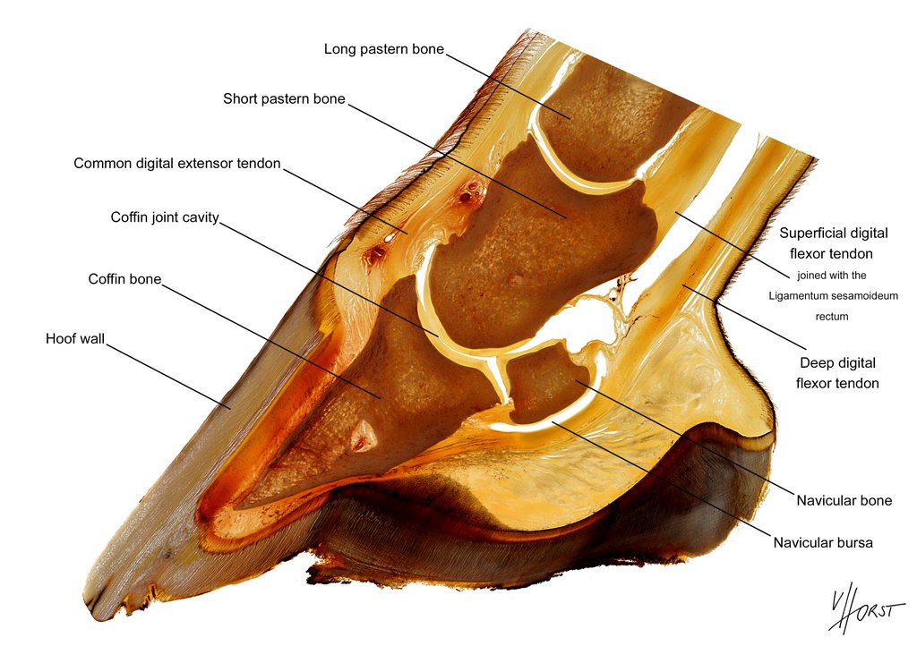
2. Gross anatomy of the equine hoof. The distal extremities of the domestic mammal are encased inside a keratinised capsule [], which takes the form of a hoof capsule in ungulates and a claw in carnivores [].This insensitive horny structure encloses the distal part of the second phalanx (also known as the middle phalanx or short pastern bone), the distal phalanx (also known as the coffin bone.
Labeled Hoof Diagram Cavallo Hoof Boots Horse Boots
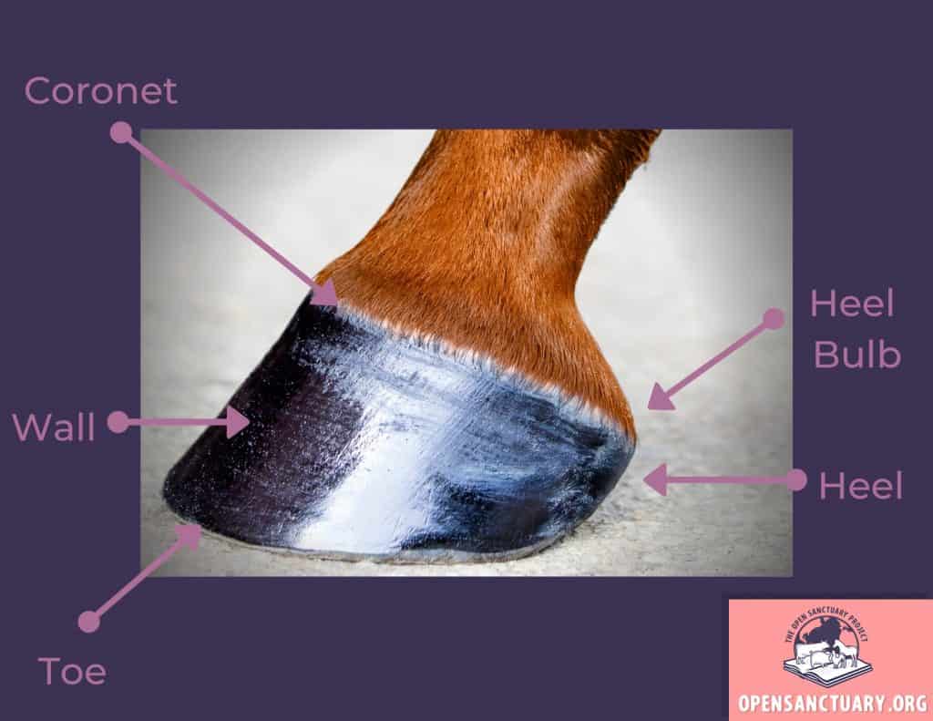
General Anatomy Of The Hoof. Let's start by looking at the following diagram, which shows basic outer hoof anatomy. Knowing these words and the areas they refer to on a horse's hooves will allow you to better understand your resident's mobility, provide better care, and communicate more effectively with an equine veterinarian and farrier.
FileHorse anatomy hooves.jpg Wikipedia
Hoof Wall. The first part of the hoof that you'll notice is the hoof wall. This is the hard, pigmented outer layer that houses and protects the more delicate structures within. Its purpose is to support the horse's weight, absorb shock as it moves, and is the first line of defence against injury and disease. The hoof wall is made up of a.
Horse hoof anatomy teaching chart Plastination Anatomy Embedding
Edit, Fill & eSign PDF Documents Online. No Downloads Needed. Get Started Now. Best PDF Fillable Form Builder. Professional Toolset. Quick and Simple. Subscribe for more
Horse Hoof Anatomy A Guided Tour The Horse
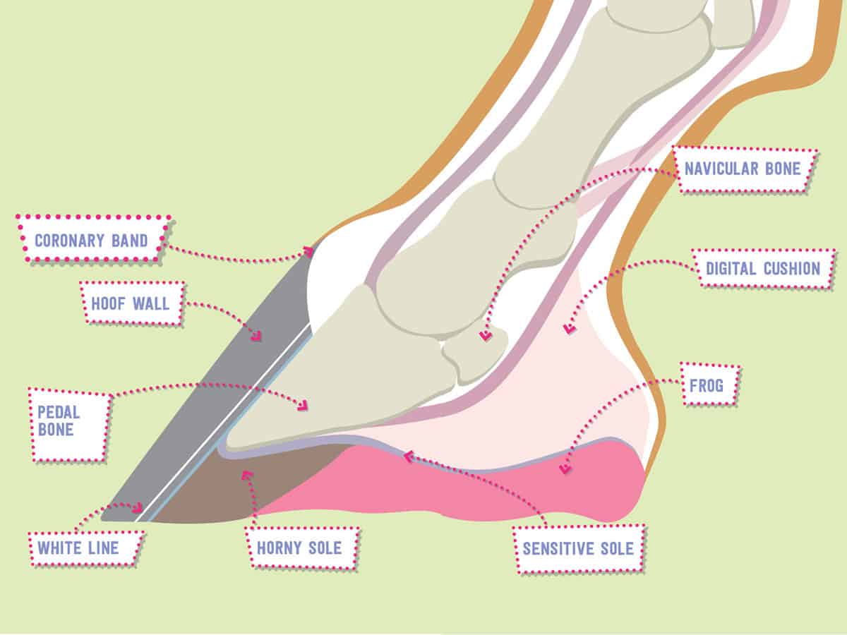
Coffin Bone. The coffin (or "pedal") bone is the bottom bone located near the toe and encapsulated in the hoof. It is the largest bone in the hoof and helps to shape the hoof wall. It's surrounded by special tissues that help make-up the laminae of the hoof wall, as well as, the tissues of the sole.

Horse Anatomy The Hoof The Open Sanctuary Project
Function 3: Communication. Every step reveals critical information the horse must know, says Catrin Rutland, PhD, PGCHE, MMedSci, SFHEA, FAS, associate professor of anatomy and developmental.

Hoof anatomy Horse care, Horse anatomy, Anatomy
Hoof Anatomy. This page shows the b asic external hoof anatomy with all the landmarks clearly labeled. These photos will help you visualize everything inside of the horse's hoof, understand the relationship between the parts and learn to read the clues the hooves have to offer. Horses hooves are amazing structures.
Horse Anatomy The Hoof The Open Sanctuary Project
Inflammation of the sensitive laminae which attach the hoof capsule to the fleshy portion of the foot. In laminitis, the blood flow to the laminae is affected, resulting in inflammation and swelling in the tissues within the hoof, and severe pain. As the laminae are starved of oxygen and nutrient rich blood, the cells become damaged.

hoof Kids Britannica Kids Homework Help
The anatomy of the equine hoof can be intimidating, but the hoof can be broken down into three groups to make it easier to understand. Anatomy of a Horse's Hoof Inner Structures Digital Cushion. The digital cushion is a mass of flexible material that lies below the coffin bone.
Hoofanatomydiagram Pony Magazine
The digital cushion of horse hoof anatomy. The digital cushion of horse hoof anatomy is a wedge-shaped mass that overlies the frog. You will find four different surfaces, a base, and an apex in the digital cushion of a horse hoof. The deep surface of the digital cushion of horse hoof faces upward and forwards.
The Anatomy, Histology and Physiology of the Healthy and Lame Equine
Hoof Anatomy - A Beginner's Guide. The horse's hoof is a miracle of engineering. It contains a whole host of structures which, when healthy, operate in equilibrium with each other to form a hoof capsule which is able to withstand huge forces, utilising energy to assist with forward movement while providing protection to the sensitive.

HOOFsmart · Hoof Anatomy Cross section photo
How the Hoof Fits Into the Anatomy and Physiology of the Horse: The best place to start is with a basic understanding of how the hoof fits into the anatomy and physiology of the horse. The largest organ (glandular structure) of the horse is the dermal tissue, a voracious consumer of nutrients which includes not only the hooves, but also the skin, hair follicles, sweat glands, oil glands and.